The cardiovascular system, often called the circulatory system, acts as the body’s lifeline, ensuring every cell gets what it needs to survive. The cardiovascular system is made up of three key components: the heart, blood vessels (veins, arteries, and capillaries), and blood.
The heart’s main purpose is to pump blood throughout the body to supply cells and tissues with the food and oxygen they need to survive. The body contains approximately six quarts of blood. By completely circulating all six quarts through the body every one to three minutes, the heart keeps the living system—our body—running. Artery carry blood to the body from the hear and veins carry blood to the heart from the body. Right side of the heart carries deoxygenated blood, whereas the left side of the heart carries the oxygenated blood.
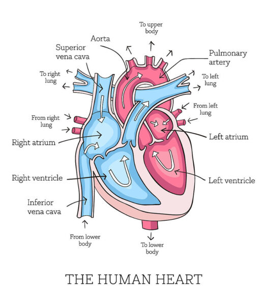
The different components of a heart are, it is divided into 4 chambers:
- Septum
- Partition between the 2 ventricles
- Interventricular septum is a muscular wall that separates right and left ventricles
- It prevents the mixing of oxygenated and deoxygenated blood
- To separate two atrial chambres so that there is no shunting of blood between them
- Vena Cava
- Superior – Blood flow from above the heart
- Inferior – Blood flow from below the heart
- Largest bein in the body
- Right Atrium
- Receives blood from body
- Empties the blood into the right ventricle when full
- Right Ventricle
- Receives blood from RA and send to the pulmonary artery
- Left Atrium
- Receives blood from lungs through the pulmonary veins
- Empties the blood into the left ventricle when full
- Left ventricle
- Receives blood from LA and send to the Aorta
- Pulmonary artery
- Connected to the lungs, deoxygenated blood
- Pulmonary vein
- Carrying oxygenated blood from the lungs to the left atria of the heart
- Pulmonary valves
- Helps the blood flowing in the correct direction through the heart
- Mitral valve
- Bicuspid valve
- Tricuspid valve
- Remains closed until blood pressure is full, when the aorta is filled. Artium’s are filled
- Semilunar valves
- Mitral valve – Making sure the blood flows from your left atrium to your left ventricle. Prevents backflow of blood into the atrium.
- Tricuspid valves – Allowing blood flow into the heart from the body to the right ventricle where it’s pumped to the lungs for oxygen.
- Pulmonary valve controls the flow of blood between heart and lungs
- Aortic controls the flow between heart and parts of the body
- The opening and closing of these valves leads to your heartbeat
- Aorta
- Main artery of the heart
- Takes blood from LV and transports throughout the body
The cardiac cycle is a coordinated sequence of contractions and relaxations by the heart muscle which cause blood to move from the atria, into the ventricles and then the arteries.
Systemic Circulation
Systemic circulation moves blood between the heart and the rest of the body. It sends oxygenated blood out to the cells and returns deoxygenated blood to the heart.
Pulmonary Circulation
Pulmonary circulation moves blood between the heart and the lungs. It transports deoxygenated blood to the lungs to absorb oxygen and release carbon dioxide. The oxygenated blood then flows back to the heart.
Oxygenated blood
- A blood cell with high percentage of oxygen and a low percentage of carbon dioxide is oxygenated blood
- Carried by arteries
- Oxygenated blood puts more pressure on arteries.
Deoxygenated blood
- Deoxygenated blood is the blood that has low oxygen saturation in comparison to blood leaving the lungs.
- It is carried by veins
- Deoxygenated blood puts less pressure on the veins
Blood vessels
| Arteries | Veins | Capillaries | |
| Function | → Carry blood away from the heart at high-pressure | →Return blood to the heart at low-pressure | → Supply all cells with their requirements→ Take away waste products |
| Pressure | High Pressure | Low Pressure | Low Pressure |
| Structure of wall | → Thick, strong→ Contain muscles, elastic fibers and fibrous tissue. | → Thin→ Mainly fibrous tissue→ Contain far less muscle and elastic tissue than arteries | → Very thin→ Only one cell thick |
| Lumen | → Narrow→ Varies with a heartbeat (increases as a pulse of blood passes through. | → Wide | → Very narrow→ Just wide enough for a red blood cell to pass through |
| Valves | Not present | Present to prevent backflow | Not present |
| How structure fits function | → Strength and elasticity needed to withstand the pulsing of the blood, prevent bursting and maintain pressure waves.→ Helps to maintain high blood pressure, preventing blood from flowing backwards. | → No need for strong walls, as most of the blood pressure has been lost.→ Wide lumen offers less resistance to blood flow. | → No need for strong walls, as most of the blood pressure has been lost.→ Thin walls and narrow lumen bring blood into close contact with body tissue, allowing diffusion of materials between capillary and surrounding tissues.→ White blood cells can squeeze between cells of the wall. |
The cardiovascular system is truly the body’s lifeline. With every heartbeat, it sustains us, fuels our energy, and keeps us connected from head to toe.

Leave a comment